The cutaneous innervation of the lower limb describes the nerve supply to specific areas of the skin of the lower limb.
There are two distinct innervation patterns:
- Peripheral nerve pattern – area of skin supplied by a specific peripheral nerve.
- Dermatome pattern – area of skin supplied by a specific spinal nerve.
In this article, we shall look at the anatomy of both the peripheral nerve and dermatome patterns of cutaneous innervation of the lower limb.
Premium Feature
3D Model
Gluteal Region
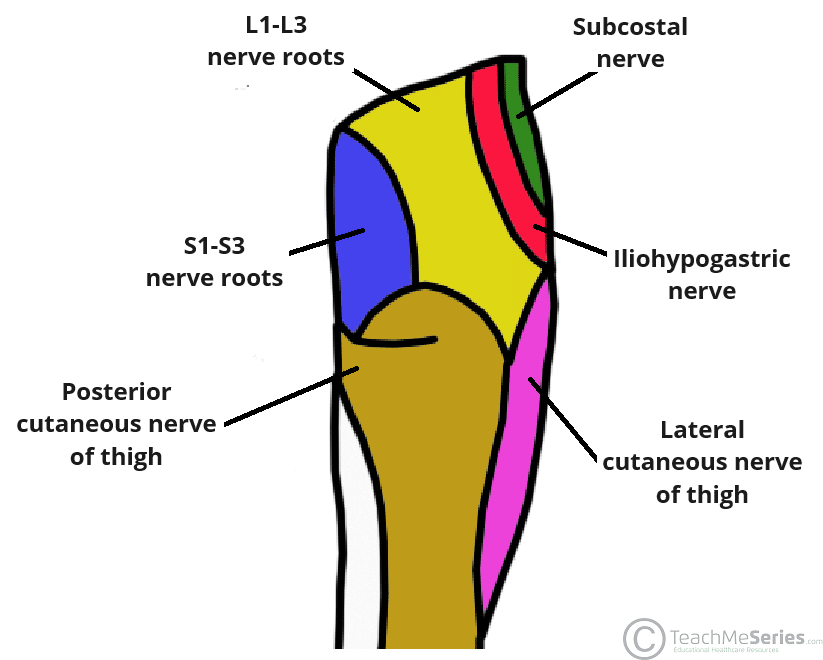
The cutaneous innervation to the gluteal region can be organised into quadrants. The skin of each quadrant is innervated by the following peripheral nerves:
- Upper medial quadrant – supplied by the posterior rami of L1-3 and S1-3 spinal roots.
- Upper lateral quadrant – supplied by the subcostal nerve (anterior ramus of T12) and iliohypogastric nerve (branch of the lumbar plexus).
- Lower medial quadrant – supplied by the posterior cutaneous nerve of thigh (branch of the sacral plexus).
- Lower lateral quadrant – supplied by the lateral cutaneous nerve of thigh (branch of the lumbar plexus)
Premium Feature
Dissection Images

Thigh Region
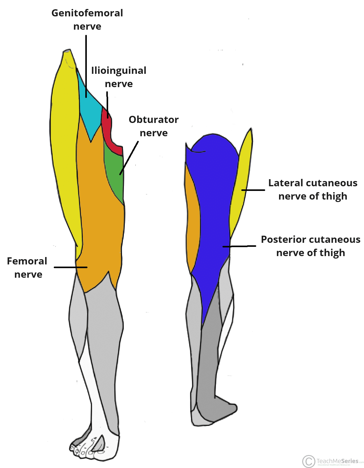
The thigh region receives cutaneous innervation from three main nerves (femoral, posterior cutaneous and lateral cutaneous). The superomedial aspect of the thigh also has contributions from several smaller branches of the lumbar plexus.
- Femoral nerve – arises from the lumbar plexus. It gives rise to anterior, medial and intermediate branches which supply the majority of the skin of the anterior thigh.
- Posterior cutaneous nerve of thigh – supplies the lower medial quadrant of the gluteal region, posterior aspect of the thigh and knee.
- Lateral cutaneous nerve of thigh – supplies the lower lateral quadrant of the gluteal region, lateral aspect of the thigh and knee.
- Femoral branch of the genitofemoral nerve – arises from the lumbar plexus. It supplies the skin overlying the lateral aspect of the femoral triangle.
- Ilioinguinal nerve – arises from the lumbar plexus. It supplies the skin overlying the medial aspect of the femoral triangle.
- Obturator nerve – arises from the lumbar plexus. Branches from the anterior branch of the obturator nerve supply skin over the medial aspect of the thigh.
Leg
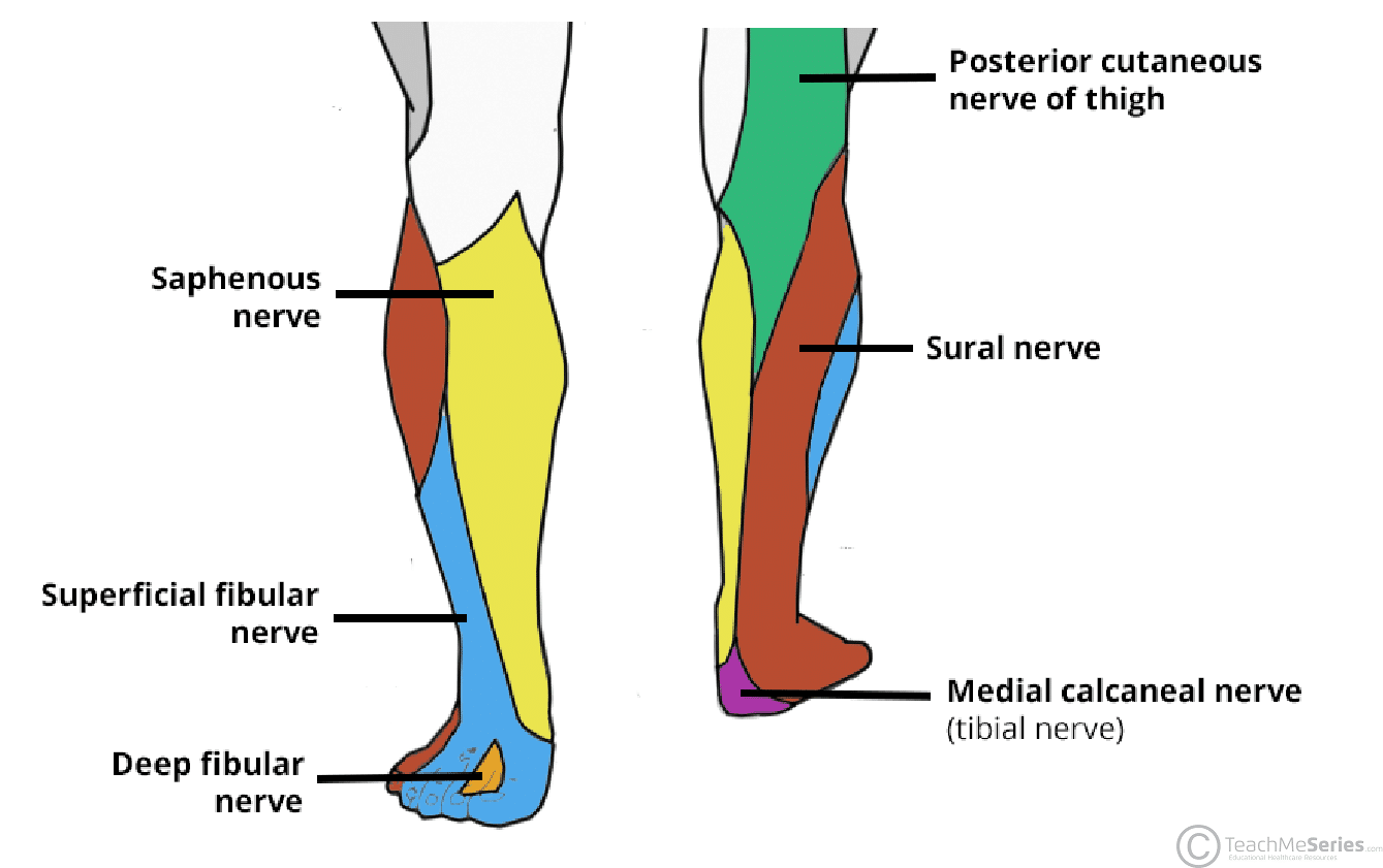
The cutaneous innervation to the leg is from the following peripheral nerves:
- Posterior cutaneous nerve of thigh – supplies the skin over the central superior and posterior leg.
- Saphenous nerve – a sensory branch of the femoral nerve. It supplies the skin over the anteromedial aspect of the leg and medial aspect of the foot.
- Superficial fibular nerve – a branch of the common fibular nerve. It supplies the skin over the anterolateral aspect of the leg and dorsum of the foot.
- Sural nerve – formed by contributions from the tibial and common fibular nerves. It supplies the posterolateral aspect of the leg and lateral margin of the foot.
Ankle and Foot
The ankle and foot receive cutaneous innervation from branches of the tibial nerve, common fibular nerve and femoral nerve:
- Deep fibular nerve – supplies the space between 1st and 2nd toes on the dorsum of the foot.
- Superficial fibular nerve – supplies the dorsum of the foot (except the space between the 1st and 2nd toes).
- Medial and lateral plantar nerves – terminal branches of the tibial nerve. The medial plantar nerve supplies the medial two thirds of the sole of the foot. The lateral plantar nerve supplies the lateral third.
- Medial calcaneal nerve – branch of the tibial nerve. It supplies the medial aspect of the heel.
- Sural nerve – formed by contributions from the tibial and common fibular nerves. It supplies the lateral margin of the hindfoot and midfoot.
- Saphenous nerve – branch of the femoral nerve. It supplies the medial margin of the hindfoot and midfoot.
Dermatomes
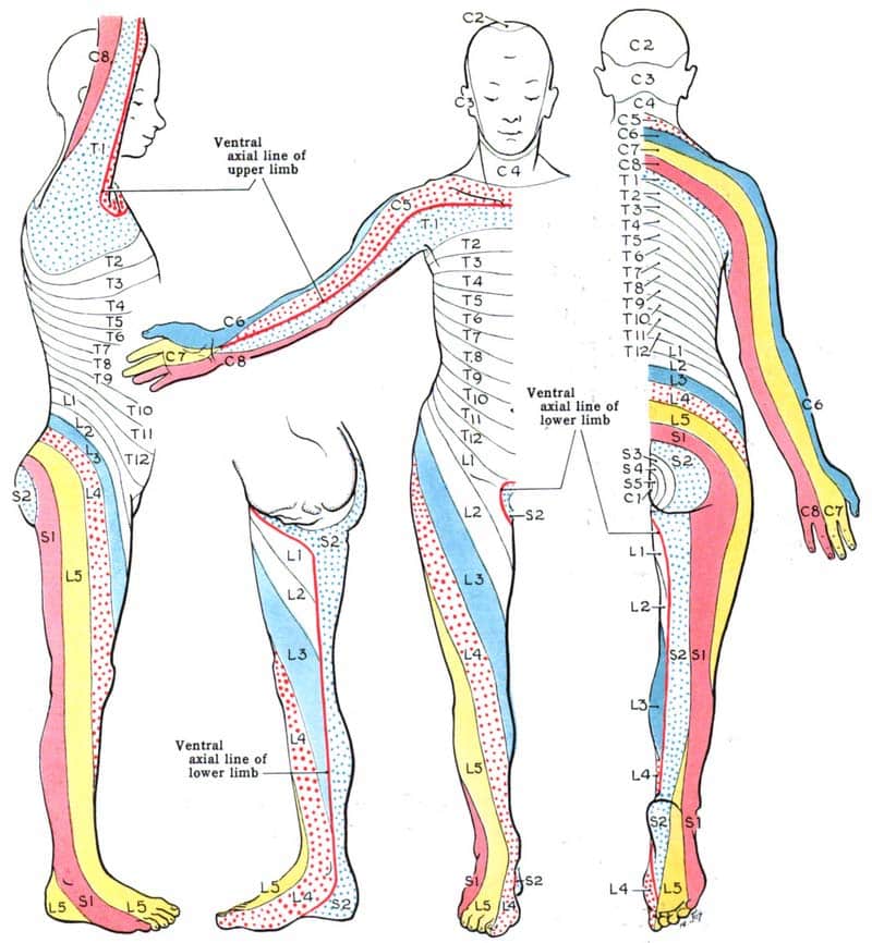
Dermatomes refer to a region of skin supplied by nerve fibres from a spinal root. They are most commonly used when assessing for spinal cord pathology
The dermatomes of the lower limb are as follows:
- L1 – Inguinal region and superior aspect of the medial thigh.
- L2 – Middle/lateral anterior thigh
- L3 – Medial condyle of the femur running inferomedial across the thigh.
- L4 – Medial malleolus (medial bony prominence of the ankle)
- L5 – Dorsum of the foot at the third metatarsophalangeal joint.
- S1 – Lateral aspect of the calcaneus.
- S2 – Midpoint of the popliteal fossa.
- S3 – Horizontal gluteal crease
- S4/5 – Perianal region


