Directory

Inguinal (Hesselbach's) Triangle - Borders - Direct Hernia - TeachMeAnatomy

The Inguinal (Hesselbach’s) Triangle

Written by Oliver Jones

Last updated August 15, 2018 • 14 Revisions •

The inguinal triangle (Hesselbach’s triangle) is a region in the anterior abdominal wall. It is alternatively known as the medial inguinal fossa.

It was first described by Frank Hesselbach, a German surgeon and anatomist, in 1806.

In this article, we shall look at the anatomy of the inguinal triangle – its borders, contents and clinical relevance.

Premium Feature

3D Model

Pro Feature
Access this feature with pro.
Go Premium

Borders

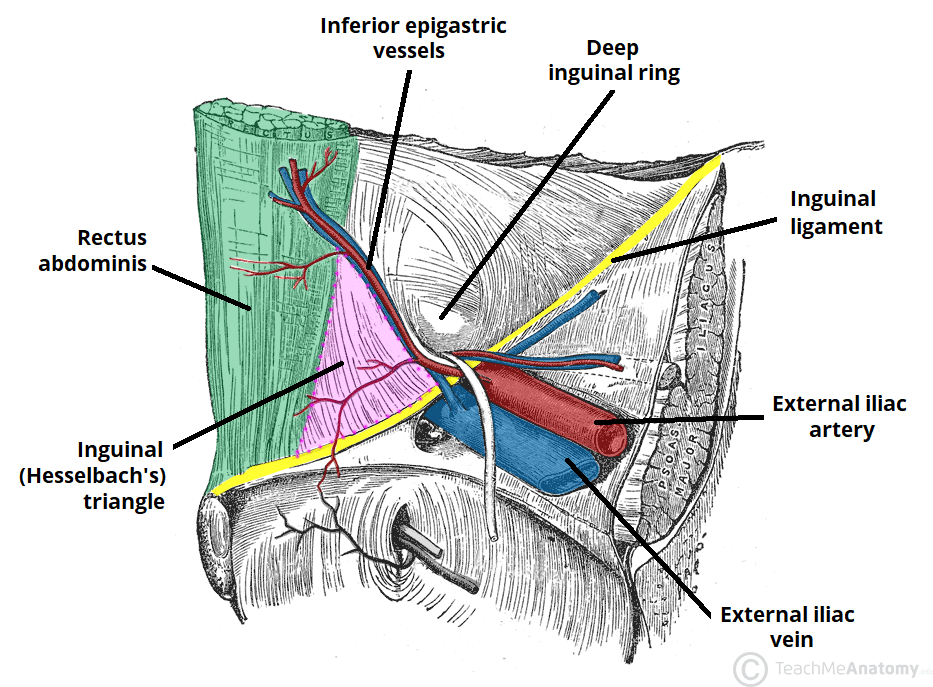
The inguinal triangle is located within the inferomedial aspect of the abdominal wall. It has the following boundaries:

  • Medial – lateral border of the rectus abdominis muscle.
  • Lateral – inferior epigastric vessels.
  • Inferior – inguinal ligament.

Contents

Other than the layers of the abdominal wall, the inguinal triangle does not contain any structures of clinical importance.

However, the triangle does demarcate an area of potential weakness in the abdominal wall – through which herniation of the abdominal contents can occur.

Fig 1
The borders of the inguinal triangle.

Clinical Relevance

Direct Inguinal Hernia

A hernia is defined as the protrusion of an organ or fascia through the wall of a cavity that normally contains it. The inguinal triangle represents an area of potential weakness in the abdominal wall, through which herniation can occur.

In a direct inguinal hernia, bowel herniates through a weakness in the inguinal triangle, and enters the inguinal canal. Bowel can then exit the canal via the superficial inguinal ring and form a ‘lump’ in the scrotum or labia majora. Direct hernias are acquired (usually in adulthood), due to weakening in the abdominal musculature.

This is in contrast to an indirect inguinal hernia – where bowel enters the inguinal canal via the deep inguinal ring.

By definition, a direct inguinal hernia occurs medially to the inferior epigastric vessels (through the inguinal triangle), and an indirect hernia occurs laterally to these vessels.

See here for more information on direct and indirect inguinal hernia.

Fig 2
Transverse view of the inferior abdominal cavity. Compares normal anatomy with indirect and direct herniation.

Do you think you’re ready? Take the quiz below

Premium Feature

Quiz

The Inguinal (Hesselbach’s) Triangle

Question 1 of 3

Submitting...
Rate question:
You scored
0%
Skipped: 0/3
Make sure you're ready by unlocking all available questions.
Go Premium
'); }); } jQuery('#submitAnswer').on('click', function(e) { e.preventDefault(); if(jQuery(this).hasClass('continue')) { jQuery(this).removeClass('continue'); jQuery('html,body').animate({scrollTop: jQuery('#quiz').offset().top}, 250); if(currentQuestion Please select an answer
'); return; } jQuery('#questionAnswers .alert').remove(); jQuery('#quizButtons').addClass('d-none'); jQuery('#quizLoading').removeClass('d-none'); jQuery.post(ajaxurl, { action: 'log_quiz_answer', id: quizQuestions[currentQuestion - 1].question_id, answer: jQuery('input[name="quizAnswers"]:checked').val(), type: 'article' }, function(data) { if(data.correct) correctQuestions++; jQuery('#questionAnswers').prop('disabled', true); jQuery('#quizLoading').addClass('d-none'); jQuery('#skipQuestion').addClass('d-none'); jQuery('#quizButtons').removeClass('d-none'); jQuery('#submitAnswer').addClass('continue'); let total = 0; for (i = 0; i div').each(function(i) { let correct = false; if(jQuery(this).find('input').val() == data.correctAnswer) correct = true; let percentage = (data.counts[i] / total) * 100; if(jQuery(this).find('input').is(':checked')) jQuery(this).append('
'); else if(correct) { jQuery(this).append('
'); } jQuery(this).append('
' + Math.round(percentage) + '% Answered ' + (correct ? 'Correctly' : 'Incorrectly') + '
'); }); let explanation = ''; explanation += '' + Math.round((data.counts[data.correctAnswer - 1] / total) * 100) + '% of users answered correctly.'; if (data.explanation.length > 0) explanation += '
' + data.explanation + '
'; jQuery('#questionAnswers').append('
' + explanation + '
'); if(currentQuestion >= quizQuestions.length) { jQuery('#submitAnswer').text('Finish'); } }, 'json'); }); function showResults() { jQuery('#quizQuestionsArea').addClass('d-none'); jQuery('#quizResultsArea').removeClass('d-none'); let score = Math.round((correctQuestions / currentQuestion) * 100); jQuery('#quizResult').text(score + '%'); jQuery('#skippedQuestions').text(skippedQuestions); } jQuery('#skipQuestion').on('click', function(e) { e.preventDefault(); skippedQuestions++; jQuery('html,body').animate({scrollTop: jQuery('#quiz').offset().top}, 250); if(currentQuestion
TeachMeAnatomy
Part of the TeachMe Series

The medical information on this site is provided as an information resource only, and is not to be used or relied on for any diagnostic or treatment purposes. This information is intended for medical education, and does not create any doctor-patient relationship, and should not be used as a substitute for professional diagnosis and treatment. By visiting this site you agree to the foregoing terms and conditions. If you do not agree to the foregoing terms and conditions, you should not enter this site.

© TeachMe Series 2025
Registered in England & Wales